
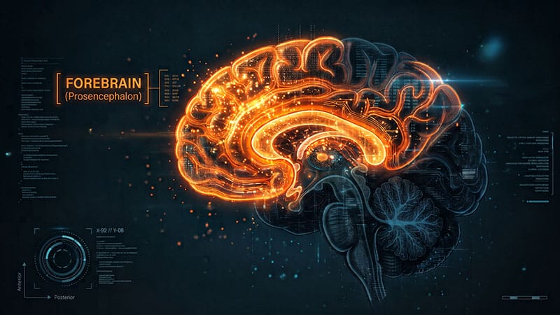
Many of us breathe unconsciously and automatically, unaware of each inhale and exhale. While breathing is fundamental to life itself, there’s still so much we do not yet know about how our brains monitor it.To be more specific, how our forebrain interprets internal breathing signals has long been a mystery: Some patients fail to recognize life-threatening shortness of breath caused by asthma, opioid use, and infections such as COVID pneumonia, while other people can experience persistent breathlessness with lungs that appear healthy and function as normal.In a recent study led by researchers from the Feinstein Institutes for Medical Research in Manhasset, New York, the team discovered a dynamic brain circuit deep within the anterior insular cortex (AIC) that can act as an “internal alarm system” and rapidly detect breathing sensation mismatches, or when inhaling suddenly feels harder than expected, for example.Jose L. Herrero-Rubio, PhD“In earlier research, we found that breathing synchronizes activity across widespread cortical and limbic networks and that this relationship changes when people deliberately control or attend to their breathing,” said Jose L. Herrero-Rubio, PhD, assistant professor of human brain mapping and bioelectronic medicine, and lead researcher of this study. “This led us to ask how the brain detects an unexpected breathing restriction in real time.”How They Did ItThe Feinstein team had the rare opportunity to record the intracranial brain activity and respiratory physiology from eight neurosurgical patients who already had electrodes implanted across interoceptive regions of their brains as part of their ongoing monitoring and treatment for epilepsy.During each trial, patients wore a nose clip and breathed through a mouthpiece. As they inhaled, the researchers applied brief, mild, and unpredictable inspiratory resistances, or loads. Afterward, patients reported whether they detected a load, when it occurred, and how intense the load felt. This allowed the researchers to directly link the patients’ brain activity to their conscious perception of their breathing.“We used standardized flow-resistive loads ranging from 1 to 15 centimeters of water per liter per second, delivered through the same type of calibrated breathing circuit and nonrebreathing valve,” said Herrero-Rubio.Compared with the preloaded control breaths, the inspiratory loads reduced airflow by 22.3% and increased mouth pressure by 121.1% for all patients. The magnitudes of the loads spanned values above and below individual perceptual thresholds, while respiratory rate and tidal volume remained consistent across trials. Those with lower perceptual thresholds were associated with higher detection accuracy.Participants followed visual cues that paced each inhalation and exhalation, while inline sensors continuously measured mouth pressure and airflow, allowing researchers to verify the mechanical effect of every load rather than relying only on its nominal value. “Some participants were able to detect smaller loads than others, which determined the individual perceptual threshold, which was considered for the data analyses for each participant,” said Herrero-Rubio.The researchers observed the insular and frontal cortices to be the most load-responsive sites. Across all patients, the inspiratory loads reliably increased their high-frequency neural activity relative to the preloaded control inhalations in 18% of all electrodes.The AIC notably had the highest proportion of responsive electrodes and the strongest gains in high-frequency activity (HFA). An example AIC electrode showed a 16.8% HFA increase, with effects beginning about 285 ms after inhalation onset and lasting about 1.5 s. In AIC electrodes, HFA responses were also larger during detected vs missed trials. In other words, the responses were substantially greater when participants consciously detected the resistance as opposed to when they experienced a load but failed to detect it.“We measured how closely high-frequency activity in the anterior insula aligned with the phase of the respiratory cycle,” said Herrero-Rubio. “This alignment was stronger when participants detected a load than when they missed a load of the same magnitude, showing that awareness depended not simply on the strength of the obstruction but on how effectively the brain coordinated its response with the incoming respiratory signal from the lungs.”Testing Which Part of the Brain Detects Certain StimulusTo test whether the AIC initiated load detection while frontal regions supported downstream processing, the Feinstein team compared load-evoked HFA dynamics across the brain. The electrodes exhibited transient responses in the AIC and more sustained activity in the premotor and orbitofrontal cortex (OFC) regions.To account for variability in inhalation duration, they normalized neural activity to the respiratory phase cycle and binned HFA accordingly. “We divided the respiratory cycle into 18 phase bins and calculated the average high-frequency neural amplitude in each bin,” said Herrero-Rubio. “If neural activity was unrelated to the breathing phase, its amplitude would be distributed evenly; the Kullback-Leibler-based modulation index measured how far the observed distribution departed from that uniform pattern.”“A larger modulation index therefore meant that anterior-insula activity was more tightly locked to a particular part of the breath. Comparing load-matched detected and missed trials showed stronger phase-amplitude coupling during conscious detection,” he continued.AIC responses were restricted to early-phase bins, while the premotor and OFC regions, on the other hand, remained active throughout the inhalation. Across the AIC phase cycle, HFA declined by 12% but only by 3% in the OFC. This decline was consistent with a temporal shift starting from the AIC for early detection to prolonged processing in the frontal cortices.“The cortex did not represent a breathing obstruction as one undifferentiated signal. The anterior insula primarily tracked the early rise in inspiratory pressure, frontal-motor regions tracked pressure and airflow throughout the breath, and the orbitofrontal cortex shifted from early pressure-related activity to later airflow-related activity,” said Herrero-Rubio.“This division resembles the specialization found in brainstem respiratory circuits, where some regions track airflow and timing (eg, active vs passive inspiration vs gaps), while others track effort or pressure,” he explained. “The parallel is important because it suggests that respiratory information remains organized by function as it moves from automatic control systems into the cortical networks that create awareness and guide action.”The AIC’s early, fast, and short-lived responses helped signal the initial breathing mismatch, helping patients detect the load. The sustained activity in the frontal and motor regions of the brain helped evaluate the load’s intensity, alerting the patients to how they should compensate for their sudden difficulty in breathing, such as by breathing harder.“Breathing awareness is not produced by the lungs or brainstem alone,” said Herrero-Rubio. “The forebrain actively interprets each breath: The anterior insula detects when something is wrong, then sends that information to frontal regions that evaluate the threat and organize a response.”What the Findings May MeanBy identifying a core circuit that can detect a mechanical breathing disruption, evaluate its significance, and help the body respond accordingly, the Feinstein team is hopeful that future studies can test how expectation and arousal can alter specific parts of this circuit in conditions such as persistent dyspnea, asthma, chronic obstructive pulmonary disease, long COVID, and anxiety-related breathing disorders.Muhammad Qasim, MD“This work could eventually help us understand why respiratory challenges trigger strong arousal in some circumstances but are inadequately detected in others,” said Muhammad Qasim, MD, pulmonologist and respiratory medicine specialist who was not involved in the study. “Connections to sudden infant death syndrome (SIDS), panic disorder, or opioid-induced respiratory depression are biologically interesting, but they remain hypotheses rather than findings demonstrated by this study.”Nonetheless, he underscored the relevance of this study with his work with sleep-related breathing disorders. “Clinically, it reinforces something we already see in sleep medicine: The severity of a breathing disturbance and the patient’s awareness of it are not always proportional,” said Qasim. He’d like to see if the same pathway operates during sleep and that differences in its function predict arousal, oxygen drops, symptoms, or treatment responses in people with sleep apnea.“What I find most fascinating is the distinction the investigators make between the brain detecting a change in breathing and our becoming consciously aware of that change,” said neurologist and clinical neurophysiologist Mill Etienne, MD, MPH (he was also not involved in the study). “What makes this study unusual is that the investigators had the opportunity to record directly from the brains of patients who already had intracranial electrodes implanted. That gave them millisecond-level information about what different cortical regions were doing as breathing became more difficult. So rather than identifying a single ‘breathing awareness center,’ this study suggests something more interesting to me: a network and a sequence characterized as detection, appraisal, and response.”Brian J. Dlouhy, MD“Our work has implicated interactions between the amygdala and anterior insula as one potential mechanism by which awareness of the loss of breathing may be disrupted,” said Brian J. Dlouhy, MD, pediatric neurosurgeon and epilepsy surgeon at the University of Iowa Health Care in Iowa City. “That makes the current study particularly interesting because it provides additional evidence that the anterior insula may be an important part of the brain’s system for detecting and becoming aware of changes in breathing.”What Further Work Could IlluminateDespite how promising this discovery made by the Feinstein researchers is, the outside experts note the study’s limitations, such as the size (11 patients participating while eight were included in the main analysis) and the circumstances.Mill Etienne, MD, MPH“These also weren’t healthy volunteers randomly selected from the population,” said Etienne. “They were patients with drug-resistant epilepsy undergoing invasive intracranial monitoring, and electrode placement was determined by clinical necessity. Therefore, not every participant had electrodes in every brain region the investigators might ideally have wanted to examine.”Etienne suggests the work is an important mechanistic study that provides a compelling model and generates important hypotheses, “rather than a definitive map of human breathing awareness.”Rebecca Spencer, PhD“There is an essential next step to this research,” said Rebecca Spencer, PhD, neuroscientist at the University of Massachusetts Amherst. “Translating this basic finding — where is this controlled — into what it means in cases where this detection system may go awry, such as sleep apnea. We don’t know if that is connected yet or how this ‘cognitive control’ of breathing may come into play in breathing disruptions and disorders. But hopefully this [finding] motivates that work.”Etienne agreed. “Studies like this one remind us how much we still don’t understand,” he said. “We breathe thousands of times every day, mostly without thinking about it. Yet we are still trying to understand how the brain takes those physiologic signals, makes us consciously aware of them, and determines how we respond.”Spencer, Dlouhy, and Etienne reported having no conflicts. Disclosure information for study authors is available in the original study publications.
Solving the Mystery of How Our Brain Allows Us to Breathe
Full Article
Original Source
Read the full article at Medscape →KhanList aggregates and links to publicly available news content. We do not host full articles from third-party sources. Always verify important information with original sources.