
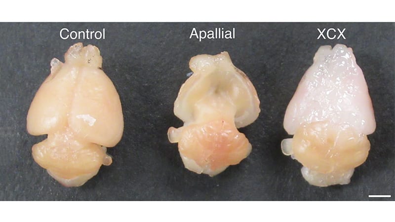
Newly developed “apallial” mice are forgetful, have a cautious gait, have notable behavioral variation from one mouse to the next, and are missing 98% of their cerebral cortex and hippocampus.The sprawling open space in their tiny skulls is intentional.Sergiu Pasca, MDNeuroscientist Sergiu Pasca, MD, and his team developed the mice to receive human cortical organoid transplants. The Stanford University professor calls the process “xenocortication.”And it works.Named for their lack of dorsal and medial palliums, the 2-day-old mice received up to four human stem-cell-derived cortical organoid transplants that integrated into the central nervous system and improved gait and maze abilities over a 3-month period. Their brains grew nearly fivefold.A hypoxia experiment produced intriguing results, and the transplanted organoids gave rise to a rare nerve cell type that scientists haven’t been able to produce in culture.Xenocortication is just the latest waymark Pasca has stamped on his quest to access human brain development. First, he worked on patient-derived cells, then three-dimensional organoids. Next: assembloids. In 2022, he reported that brain organoids transplanted into rats resulted in rat cells migrating to the human tissue. Now, his team has integrated large-scale human cortical tissue into a living nervous system.After each accomplishment, Pasca says the same thing: His focus is neuropsychiatric conditions, and the goal is intervention and therapeutics. He’s just on the trail to access the underlying biology of human nervous system development.“For me, the motivation has always been disease,” Pasca said. “By the time a patient comes to the clinic, we are generally seeing the consequences of those developmental events rather than the events themselves.”“Building models is not the endpoint” and “the intent is not to develop transplantation itself as a treatment,” he said. “The purpose of the models is to make previously inaccessible biology experimentally accessible and eventually actionable.”But the results are so compelling that they’ve renewed calls for international oversight of brain organoid work.Scientists now have a new way to examine the brain’s progression from genes to cells to circuits to functions, but the unprecedented access is prompting broader ethical questions and giving rise to new scientific ones.Translational IntrigueThe transplanted organoids reached the equivalent of second trimester gestation, based on single-cell RNA sequencing, pseudobulk analysis, and benchmark comparison of glutamatergic neurons.Elizabeth Crouch, MD, PhD“That’s useful and very early for many processes,” said Elizabeth Crouch, MD, PhD, neonatologist and associate professor of pediatrics and stem cell biology at the University of California, San Francisco (she was not involved in the study). Her lab studies vasculature growth and central nervous system connections. “In humans, there is a ton of neuronal migration and synaptic development that happens in the third trimester, and then throughout the first years of life.”Among the key takeaways from the work was simply the level of neuroplasticity at this developmental stage, Pasca said.“One of the most surprising findings was that these animals were viable and, at first glance, remarkably functional,” he said. “That tells us something important about the plasticity of the developing nervous system.”In gait analysis alone, distinguishing the experimental mice from the control mice was difficult. Crouch was struck by supplemental video from the study showing the similarities.“It’s remarkable how much of the cortex is not required to have some basic functions of the mouse,” she said. “Their strategy was effective, and when they did the xenotransplantation, it seems like they captured the full range of cortical and glial classes in the xenografts.”The findings made her recall a dean at her medical school who was found to have a cyst taking up a large part of his frontal cortex as an incidental finding on a brain MRI after a fall. “He was brilliant, and he was missing, say, 10% of what the rest of us have in our cortex. This all goes to say there’s a lot to discover about neuroplasticity in terms of the elevated solve rate.”Because the hippocampus and neocortex play key roles in memory formation, the researchers examined the mice’s working memory in a Y-maze task compared with control mice and apallial mice without transplantation.The maze task “relies on the mouse’s native preference to explore novel environments, with the assumption that the mice must maintain a memory trace of the previously explored environments,” the authors wrote in the Nature paper. “Indeed, control and [transplanted] XCX, but not apallial mice, performed above chance levels. This effect was reproduced in two independent cohorts tested months apart.”Pasca’s team also analyzed an environment-based experiment: oxygen deprivation.“The developing human brain can be highly vulnerable to hypoxia, while mice are comparatively resilient to some levels of oxygen deprivation that can injure human neural tissue,” Pasca said. “In xenocortical mice, low oxygen caused substantial injury to the human cortical cells and was accompanied by abnormalities in gait and motor coordination.”The control mice were not affected, suggesting the model’s potential for study of perinatal hypoxia associated with cerebral palsy.“That gives us a way of asking why human neural cells are susceptible and whether there are interventions that could protect them,” said Pasca, who also wants to use the model to explore the impact of infection and inflammatory conditions during pregnancy.Crouch called the hypoxia experiment “particularly impactful. The mouse itself is a technical feat, and to go one step further and show its relevance for medicine, that was very nice.”Pasca said the team was surprised to find von Economo neurons during the study, which have never been grown in culture.“We still know remarkably little about these cells, including why they appear selectively vulnerable in some forms of frontotemporal dementia,” he said.With Innovation, New Problems AriseHow much of what scientists observe in this model stems from the mouse, from the human tissue, or from a new phenotype?Crouch said basic cellular architecture challenges clearly exist because fiber distributions don’t match up well across the mouse groups.“This is a really interesting problem and probably more of an engineering feat that we’re going to need to figure out in the future,” she said. “When we consider the way that the vascular cells integrate with the neural cells, we’re very much moving towards collaborating with bioengineers to create unique cellular geometries that recapitulate the processes that we want to see interact. I think an in vivo scaffold is a great substrate on which to start to build this, and there’s still a lot to do.”Moving forward, Pasca hopes to follow the process that ultimately developed PCSK9 inhibitors.“Human genetics first showed that activating mutations cause severe hypercholesterolemia, while loss-of-function variants produce very low LDL [low-density lipoprotein] cholesterol and protection from cardiovascular disease,” he said. “Access to human blood and liver biology then made it possible to connect those genetic observations to the mechanism of LDL receptor turnover and validate the target directly in human systems.”“That sequence — from human genetics to accessible human tissue, mechanism, and ultimately targeted therapies — is very much the model we aspire to for the nervous system, where access to the relevant human tissue has historically been far more limited,” Pasca said.Ultimately, he foresees his model offering a way toward testing candidate therapeutics directly on human neural tissue in vivo for both efficacy and toxicity. Atop his list of conditions to study: epilepsy and autism.The Stanford team sought extensive ethical guidance during the project, and Pasca is careful to point out that this is not a human brain in a mouse.But a couple of weeks after Nature published Pasca’s paper, the journal itself published an editorial stating, “Researchers are asking whether the animals receiving implanted brain organoids might gain any human-like properties of consciousness or cognition. How should these characteristics be measured, and what extra oversight should there be for such studies?”The editorial lauded Pasca’s ethics approach and called for the scientific community to answer the question as to whether an existing or new entity should take oversight responsibility. Independent ethics advisers should be “closely involved through the life of a research project,” the editorial stated.Meanwhile, Pasca is still committed to discovering new ways of exploration that only arise from keeping an open mind.“I would like to follow the emergence of a human neuropsychiatric disorder from its earliest developmental origins, across scales, from gene to cell to circuit to function,” he said. “If we could connect those levels experimentally, I think it could fundamentally change how we think about disease mechanisms and about when and where to intervene.”Crouch reported having no conflicts. Stanford University holds patents for the generation of cortical organoids (listing Pasca as an inventor) and a provisional patent application for transplantation of organoids (listing Pasca as an inventor). Disclosure information for study authors is available in the original study publication.
Brain Organoid Transplants and Cracking the Autism Code
Full Article
Original Source
Read the full article at Medscape →KhanList aggregates and links to publicly available news content. We do not host full articles from third-party sources. Always verify important information with original sources.