
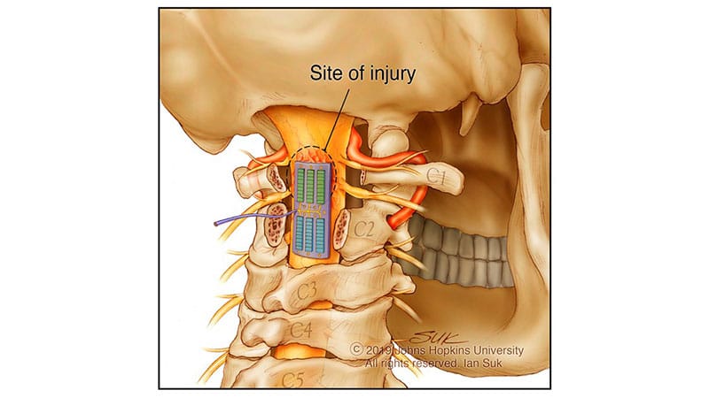
The days after decompression surgery for a spinal cord injury are a critical monitoring period for complications like hematoma and stroke, but it can be challenging to detect these issues fast enough to intervene. Even a short period of time can be long enough to cause irreversible damage. As Amir Manbachi, PhD, principal investigator and an associate professor of neurosurgery and biomedical engineering at Johns Hopkins Medicine in Baltimore, explained, “The difference between 2 hours is somebody not moving their hands.”Amir Manbachi, PhDPhysicians often monitor patients’ blood flow by measuring blood pressure, but this only provides information at the systemic level. “The injury is local,” said Manbachi. “We should locally monitor.”How to do that? Ultrasound will work but has limitations. It generally cannot observe the site of a spinal cord injury because of the thick layer of bone blocking the area. In addition, ultrasound only provides data at the time the device is used, offering no constant, on-demand feedback.An implant would solve both problems. The idea of implanting an ultrasound inside a patient’s body may sound like science fiction, but Manbachi’s team at Johns Hopkins Medicine is working on a device intended to do exactly that, collecting continuous ultrasound data from the site of a spinal cord injury as patients recover from surgery. In late October, the team published a proof-of-concept study in Science Advances.How They Did ItThe device — which they called the Multimodal Ultrasound System for Injury Care (MUSIC) — is the size of a small Band-Aid and can be implanted without interrupting surgeons’ existing workflows, said study author Joshua Doloff, PhD, associate professor of biomedical engineering at Johns Hopkins. “Because of that access that the surgeon has to take in order to even get at the tissue to intervene surgically,” he said, “it gave us the opportunity to piggyback on top of that procedure and to basically be able to place a real-time monitoring system.”The device combines both traditional diagnostic ultrasound, which monitors how the wound is healing, with the more novel therapeutic ultrasound, which responds to healing changes as they occur. With MUSIC, the therapeutic aspect happens through vasomodulation or regulation of blood flow through changes to the blood vessels. “It’s as if you’re putting ice [on it], but you don’t need the temperature to modulate,” said Manbachi.The MUSIC device delivers both diagnostic and therapeutic ultrasound directly to spinal injury sites, allowing for continuous monitoring.So far, the researchers have studied MUSIC using human cadavers and animal models, including pigs. “Their torsos and their body size and scale are more faithful to that of a human,” said Doloff. When it comes to the spinal cord, he added, “[i]t’s a faithful representation of human anatomy and human tissue response that you see in patients in the clinic.”Joshua Doloff, PhDEven so, working with pigs posed some unique challenges. For example, the researchers had to figure out the most ethical way to induce injury in the pigs so the device could be tested on them. “We actually had a lot of discussions about how can we induce enough injury to get changes to the tissue, but not cause complete deterioration of the animal,” said Doloff. “We wanted to make sure that they could still have walking ability…that their quality of life was not diminished in a significant way.” In the end, the pigs were injured in a way that mirrored a milder human spinal cord injury, meaning the pigs stayed fully ambulatory after the injury.Animals also communicate differently than humans. “Humans can have a conversation with a doctor and understand the severity of their case and know how to cooperate,” said Doloff. Because the researchers couldn’t ask the pigs if the device was causing discomfort, they had to learn about this by observing their behavior. “If [pigs] have irritation, you’ll see them rubbing,” said Doloff, but they did not observe any behaviors indicating that the pigs were uncomfortable with the device implanted.Next StepsOverall, results from the proof-of-concept study showed that the device was feasible, while testing of the device’s histologic tolerability indicated that it was safe for further testing. “[MUSIC] was not adding additional toxicity or damage to the tissue,” said Doloff.Still, it’s unclear if the therapeutic part of the technology can truly be expanded to spinal cord injury. “Therapeutic ultrasound is primarily used for musculoskeletal applications,” said Andrea Leyton-Mange, MD, a rehabilitation medicine physician at Mount Sinai Health System in New York City, who specializes in spinal cord medicine (and was not part of the study). “Musculoskeletal tissue tendons are not neural tissue, and the idea of delivering energy via an ultrasound to the injured spinal cord doesn’t have a lot of basis in existing medicine.” Because therapeutic ultrasound carries risks like cavitation, Leyton-Mange cautioned that much more research will be needed to determine if this technology is safe to use in humans.“I won’t say it’s not worth investigating,” she said. “[MUSIC] is a really cool idea. But I think the diagnostic aspect of it is probably closer to being safety tested, and in a way that would be implementable in the nearer future.”The team plans to continue studying the device and eventually bring it into trials with human patients. Although the research is still at an early stage, MUSIC has already attracted federal support, including funding from the Defense Advanced Research Projects Agency because of its potential to help soldiers who suffer spinal cord injuries. The FDA also gave the device a Breakthrough Device Designation in 2023, which is used to accelerate the development of devices that have potential to offer major clinical advantages compared with the currently available technology.If MUSIC is found safe and effective in human patients, it could help physicians make better personalized care decisions that address complications faster. “With the variety of ways in which someone can be injured, not everyone’s injury is going to look exactly the same,” said Doloff, “and so the ability to be able to make [monitoring] more personalized to the patient is incredibly powerful.”Disclosure information for study authors is available in the original study publication. Leyton-Mange reported no conflicts.
Tiny Ultrasound Implant Monitors Spinal Injury in Real Time
Full Article
Original Source
Read the full article at Medscape →KhanList aggregates and links to publicly available news content. We do not host full articles from third-party sources. Always verify important information with original sources.