
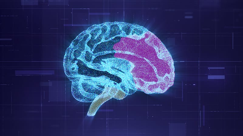
The part of your brain reading this sentence and the part keeping you breathing while you read it may have different developmental origins.That’s the idea at the center of a new study from Stanford University, published in Nature Neuroscience. The university’s press release ran with the provocative headline: “Human brain is two separate organs.”The paper itself was a bit more careful with its language. The authors postulated that the brain is a “composite organ,” built from two groups of progenitor cells that emerge in parallel during gastrulation, when an embryo begins laying out its body plan. One group goes on to form the forebrain and midbrain, and the other forms most of the hindbrain, which includes the cerebellum and the pons and medulla in the lower brainstem. The pons and medulla help control breathing and swallowing, as well as movement in the face and throat.Rayyan JokhaiRayyan Jokhai, a Stanford graduate student and co-first author of the study, compares the brain to the heart, “which is similarly constructed from at least two parallel progenitor populations,” he said. In the heart, one population primarily forms the left ventricle, while the other contributes to the remaining three chambers.The experiments support the brain’s two distinct starting points, but whether that means the regions evolved as two independent organs is a more difficult question to answer.Once the researchers saw that the hindbrain follows its own developmental path, they found a way to grow specific human hindbrain motor neurons, something stem cell labs have struggled to do. Diseases such as amyotrophic lateral sclerosis (ALS) and spinal muscular atrophy (SMA) can affect motor neurons involved in swallowing and breathing.Researchers rarely have access to brainstem tissue from living patients to study what goes wrong, but they may now be able to grow some of those neurons in a dish.The Study Began by Looking Further BackEarlier efforts to grow hindbrain neurons focused on the desired final product, then tried to reproduce the signals that would turn an early neural cell into that type of neuron.The Stanford team went further back, to gastrulation, when the embryo’s basic body plan is first taking shape. Graduate students Carolyn “C” Dundes and Jokhai, the study’s co-first authors, examined whether the hindbrain might be following its own path before recognizable brain regions had formed.Carolyn “C” DundesResearchers already knew that the genes Otx2 and Gbx2 help identify different regions of the developing nervous system. Otx2 marks the future forebrain and midbrain. Gbx2 marks cells headed for the hindbrain. “Our study suggests that precursor cells commit to a forebrain/midbrain or a hindbrain fate earlier in development than we previously thought, during gastrulation,” Dundes said.Using lineage tracing in mouse embryos, the researchers followed the descendants of the two cell populations. Cells from the anterior neural ectoderm contributed to the forebrain and midbrain, while cells from the posterior neural ectoderm contributed to the hindbrain.The team then used human pluripotent stem cells to model the two populations. In the anterior and posterior cells, different stretches of DNA were accessible, while others were packed away. That pattern was already visible before either group matured into neurons.‘Not Possible’ or Still an Open Question?Jokhai put it bluntly in Stanford’s press release. “Previous attempts to make hindbrain neurons likely tried to coax forebrain and midbrain progenitors into hindbrain cells, which our study shows is not possible.”Kyle Loh, PhDHowever, senior author Kyle Loh, PhD, associate professor of developmental biology at Stanford University School of Medicine in Stanford, California, describes that point as unresolved.“An important question left unresolved by our work is whether the two brain progenitors display any degree of plasticity,” Loh said. “Namely, whether the forebrain/midbrain progenitor could be ‘coerced’ to artificially become hindbrain, and vice versa.”Loh’s question gets at how fixed those early cell identities really are. The experiments showed the two groups developing differently under the conditions tested, but not whether different signals or a genetic change could make one group take on the other’s role. A New Source of Hindbrain Motor NeuronsThe Stanford researchers began with human pluripotent stem cells, which can develop into many kinds of cells, and guided them toward becoming hindbrain motor neurons. The lab-grown neurons had features associated with rhombomeres 5 and 6, two temporary sections of the developing hindbrain that help organize the nerve connections involved in facial movement and swallowing.The team tested whether the cells behaved like neurons. They fired action potentials, rapid electrical impulses neurons use to communicate, and made proteins typical of those hindbrain sections. The results showed that the cells had key features of neurons and the right regional markers.Both ALS and SMA can destroy these kinds of motor neurons, and SMA is a leading genetic cause of death in children younger than 1 year. Dundes hopes other labs will use the protocol to build better lab-grown models of these diseases.The study did not model ALS or SMA. Researchers plan to compare hindbrain motor neurons grown from stem-cell lines from people with SMA and unaffected controls. Dundes said, “An important next step would be to create more mature hindbrain motor neurons in vitro, which would hopefully allow us to more faithfully model these neurodegenerative diseases.”The next test is whether the cells develop changes seen in patients. If they do, researchers could use them to investigate why some motor neurons fail and test possible treatments.From Acorn Worms to JellyfishThe researchers also looked for clues to how far back the two-part pattern might go. They compared which genes were active in developing embryos from mammals, chickens, zebrafish, and acorn worms and found similar patterns across the animals.Acorn worms, small marine animals that live on or beneath the seafloor, belong to a group called hemichordates, which sits outside the chordates, the group that includes vertebrates. Finding a similar pattern in acorn worms suggests the early gene programs may be much older than the vertebrate brain. The paper proposes that they may have been conserved for roughly 550 million years, moving from hemichordates to mammals.The paper’s 550-million-year figure describes the span across which the related pattern may have been conserved, from hemichordates to mammals. Loh’s reference to chordates emerging about 500 million years ago is a different marker. He says the molecular foundation for distinguishing the progenitors was laid down before then.Loh cautions against reading too much into the comparison. “Just because these progenitors exist during early developmental stages doesn’t mean that an acorn worm has an actual hindbrain at later developmental stages, for instance,” he said.Detlev Arendt, PhD, senior scientist at the European Molecular Biology Laboratory in Heidelberg, Germany, says the evolutionary conclusion needs evidence beyond the developmental lineage. “While this is frequently a good proxy for evolutionary lineage, this is not necessarily so,” he added.Detlev Arendt, PhDArendt says the evidence extends beyond the animal group that includes acorn worms and humans. He points to annelid worms and cnidarians, including jellyfish, as groups where researchers have found evidence for a dual origin of neuroectoderm.In 2013, Arendt and Maria Antonietta Tosches published “The bilaterian forebrain: an evolutionary chimaera” in Current Opinion in Neurobiology. Bilaterians are animals with left and right sides, including humans. The authors argued that the forebrain itself combines two older systems, an apical nervous system and another on the ventral body side.While the Stanford study doesn’t cite the 2013 Tosches and Arendt paper, Arendt says it’s “very relevant” to their research and “would have strengthened their argument.” Their proposed split is in a different place. Tosches and Arendt described two systems within the forebrain, while the Stanford study places its division between the forebrain/midbrain and the hindbrain.How Could the Evolutionary Claim Be Tested?Arendt described two ways to tell whether a pattern was inherited from a common ancestor or evolved independently. One is cladistics, which compares how species are related on the evolutionary tree. If a similar pattern appears in animals separated by a long stretch of evolution, it may have evolved more than once. Scientists call that convergence.Arendt says researchers can test the evolutionary idea by comparing single-cell sequencing data from more animals. The data show which genes are active in individual cells. A close match across many genes, especially when evolution had other ways to solve the same problem, would support a shared history.The Stanford study supports two separate starting points in brain development. Whether they reflect two nervous systems that came together over evolutionary time remains an open question. Loh and his colleagues want to trace where the spinal cord fits and find out whether other organs also develop from separate groups of early cells.“Beyond the brain and heart, it would be exciting to explore if other organs are similarly built from multiple progenitor populations,” Loh said.Stanford University has filed patent applications related to neural differentiation. Loh, Dundes, Jokhai, and other study authors are listed as inventors. Disclosure information for study authors is available in the original study publication. Arendt reported having no conflicts.
Testing a Brain Theory 550 Million Years in the Making
Full Article
Original Source
Read the full article at Medscape →KhanList aggregates and links to publicly available news content. We do not host full articles from third-party sources. Always verify important information with original sources.