
TOPLINEA retrospective study found moderate or severe silent vertebral fractures in 14.6% of 2446 UK Biobank participants aged 40-79 years, with prevalence rising from 9.4% among those aged 40-49 years to 19.6% among those aged 70-79 years. Prevalence was higher among men than among women (18.8% vs 10.8%), and many fractures were seen in people with normal bone mineral density (BMD).METHODOLOGYResearchers retrospectively analysed DEXA spine images from 2446 UK Biobank participants (mean age, 62 years; 1151 men) without a known vertebral fracture or spinal trauma. Scans were obtained from 2014 to 2016 at the Stockport Clinical Research Facility in England using a GE iDXA system. Fractures were detected with an AI-assisted six-point morphometric algorithm, and the results were manually adjusted and verified by a radiologist. Moderate fractures were defined as 25%-39.9% vertebral height loss, and severe fractures were defined as 40% or greater vertebral height loss. The main outcome was fracture prevalence, and associations between fracture prevalence and age, sex, BMD, BMI, medication use, and recent back pain were assessed using multivariable logistic regression. TAKEAWAYModerate or severe vertebral fractures were identified in 14.6% of participants; prevalence was 9.4% at ages 40-49 years and 19.6% at ages 70-79 years. The prevalence of fractures was higher among men than among women (18.8% vs 10.8%; adjusted odds ratio [aOR] for women vs men, 0.51; P < .001). The odds of fracture were more than twofold higher at ages 70-79 years than at ages 40-49 years (aOR, 2.12; P = .015). Compared with participants with a normal BMI, those with obesity had higher odds of fracture (aOR, 1.99; 95% CI, 1.43-2.77; P < .001). Osteoporosis showed a significant association relative to normal BMD (aOR, 1.95; 95% CI, 1.11-3.28; P = .020), whereas osteopaenia did not. IN PRACTICE"Silent vertebral fractures are common in asymptomatic adults, are present from midlife, and are more prevalent in men. Many occur in individuals without low BMD and are not predicted by conventional clinical risk factors," the authors wrote."Radiological assessment, including opportunistic evaluation on DXA imaging, plays a key role in identifying this under-recognised condition," they added.SOURCEThe study was led by David John Wilson, University of Oxford, Oxford, UK. It was published online on October 13 in European Radiology.LIMITATIONSParticipants were UK Biobank volunteers, mostly of British ethnicity, limiting the generalisability of the findings. As the study was cross-sectional, it could not establish when fractures occurred or how they progressed, and data on symptom severity and duration were limited. Fractures were identified without an external reference standard, and agreement between the radiologists and the automated system was low. DISCLOSURESThe study received no specific funding. One author's position was supported by a Wellcome Collaborative Award. Two authors reported serving as directors of St Luke's Radiology Oxford Ltd, which provides private and outsourced NHS radiology services, and one author was affiliated with Oxehealth. This article was created using several editorial tools, including AI, as part of the process. Human editors reviewed this content before publication.
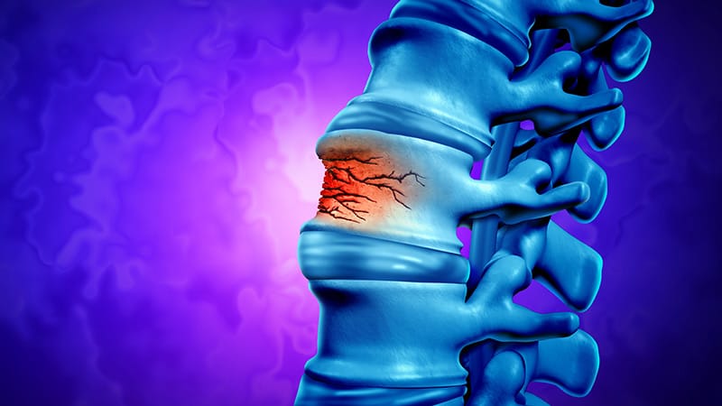
Silent Vertebral Fractures Common in Midlife, More So in Men
Full Article
Original Source
Read the full article at Medscape →KhanList aggregates and links to publicly available news content. We do not host full articles from third-party sources. Always verify important information with original sources.