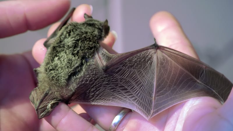
Key TakeawayExposure to a bat without a visible bite wound, nonspecific symptoms, and an initial misdiagnosis: What begins as what appears to be a herpes infection develops into fulminant encephalitis within a few days. The case published in the CMAJ of an 11-year-old boy from Canada illustrates how deceptive the course of rabies can be — and why any direct contact with a bat must be medically evaluated.The Patient and His Medical HistoryThe patient is 11 years old, immunocompetent, and had been healthy. When his parents brought him to the emergency department of a municipal hospital in Ontario, Canada, he complained of painful difficulty swallowing and vomiting. The boy had no known allergies and no tick bites and had not traveled abroad. There were no known infected contacts.The symptoms began about a week earlier: Initially, the boy felt tingling and numbness on the right side of his face. The symptoms worsened; he lost his appetite and later developed swelling on the right side of his face. Four days after the onset of symptoms, the family visited an emergency department for the first time. There, doctors suspected Bell palsy, likely associated with a herpes simplex infection. The boy was prescribed valacyclovir, 1 g three times a day. However, due to severe pain when swallowing, he was barely able to take the medication.It was only during further discussion that the parents told the doctors in the emergency room about an incident they had not previously considered dangerous: 19 days before the onset of symptoms, the family had been at a vacation home in northern Ontario. During the night, the boy woke up because a bat was perched on his nose and mouth. He reflexively swatted the animal away. His father caught the bat and released it outside.Because there were no visible bite or scratch wounds on the child’s face and the bat did not appear abnormal in the parents’ assessment, the family did not seek medical help at the time.FindingsDuring the examination in the emergency room, the boy did not appear to be seriously ill overall. His vital signs were largely within the normal range; the only notable finding was tachycardia at 134 beats/min.Neurologically, there were mild sensory disturbances in the distribution area of the right trigeminal nerve. In addition, the doctors noted mucosal ulcers on the upper and lower gums. Further neurologic and oropharyngeal examinations initially revealed no significant findings. There was no clear facial nerve palsy at this time. The facial swelling described earlier was also no longer visible.Laboratory results also did not provide a clear picture: Electrolytes, glucose, and creatinine were within the normal range. The complete blood count was unremarkable except for leukocytosis and is consistent with an inflammatory process.Initial Presumptive DiagnosisDue to the mucosal changes and neurologic symptoms, doctors initially suspected severe herpetic gingivostomatitis with Bell palsy. On the basis of the history of contact with a bat, the emergency physician notified the local health department to evaluate the need for postexposure rabies prophylaxis. However, the boy was initially discharged with a suspected diagnosis of herpetic gingivostomatitis.The next morning, his parents brought him back to the emergency department. His condition had now deteriorated significantly. He exhibited right-sided facial weakness, increasing sensory disturbances, and slurred speech. He was afebrile and hemodynamically stable. However, tachycardia was again noted, this time at 139 beats/min.Under the working diagnosis of severe herpes gingivostomatitis with Bell palsy, the boy was admitted to the hospital. He was given intravenous acyclovir and prednisone. While he was still waiting to be admitted, his condition deteriorated dramatically: He had fever of up to 39.1 °C, difficulty swallowing, confusion, and visual hallucinations.A CT scan of the head revealed no abnormalities. However, the disease progressed rapidly. By that evening, multiple cranial nerve deficits appeared, including signs of bulbar palsy. This was accompanied by pronounced hypersalivation. To protect his airway, the boy was intubated and transferred to the pediatric ICU.Further DiagnosticsIn the ICU, the history of bat exposure took center stage. The combination of direct contact to the face, sensory deficits, cranial nerve involvement, dysphagia, hypersalivation, and encephalopathy now made rabies a suspected diagnosis.However, the treatment team also considered, as part of the differential diagnosis, more common pathogens of infectious encephalitis, including the herpes simplex virus, enteroviruses, or the West Nile virus. Noninfectious causes were also taken into account, such as acute disseminated encephalomyelitis, autoimmune encephalitis, and neuromyelitis optica spectrum disorder.In parallel with general infectious disease diagnostics, samples were collected for rabies testing: serum, cerebrospinal fluid (CSF), saliva, and a skin biopsy from the neck area. Saliva and neck skin samples, in particular, were considered especially important specimens when rabies is suspected.The CSF findings were consistent with viral inflammation of the central nervous system (CNS). Lymphocytic pleocytosis was observed with leukocyte count of 272 cells/µL, 88% of which are lymphocytes. Protein was significantly elevated at 1.22 g/L, whereas glucose level was normal at 3.5 mmol/L.MRI of the brain revealed hyperintense lesions in the brainstem, particularly in the medulla oblongata. Additional changes were found in the left caudate nucleus, in cortical gray matter — with confluence in the right posterior temporal and occipital lobes — as well as in the cervical spinal cord. These findings were not specific, but they were consistent with a suspected case of rabies.On the fourth day of hospitalization, polymerase chain reaction testing of saliva samples finally confirmed the diagnosis. Further testing identified a bat-associated variant of the rabies virus. It was therefore clear that the infection was related to the nocturnal contact at the vacation home, even though there was no visible bite wound.TreatmentEven before the diagnosis was confirmed, the boy received targeted anti-infective therapy. To address both bacterial and viral causes of encephalitis, the team treated him with intravenous ceftriaxone, vancomycin, and acyclovir. After the positive rabies test, it became clear that the disease had already reached a stage at which there is no longer any established causal therapy.Treatment focused on intensive care support: securing the airway, ventilation, stabilizing circulation, and managing neurologic and autonomic complications. Nevertheless, the team was exploring experimental options.Among other things, the doctors discussed the intraventricular administration of rabies immunoglobulin via an Ommaya reservoir: a small, dome-shaped plastic reservoir implanted under the scalp. It is connected to a cerebral ventricle via a thin catheter. This would require neurosurgical intervention. Due to the invasive nature of the procedure, the lack of proven efficacy, and the rapidly progressing neurologic damage, the family, together with the treatment team, decided against this option.An experimental gene therapy using an adeno-associated viral vector designed to express a neutralizing antibody was also considered but not pursued: The therapy was not available in the short term, its efficacy has not been proven, and the boy’s clinical condition was deteriorating rapidly.Severe complications subsequently arose. The patient developed autonomic dysregulation, ventilator-associated pneumonia, and progressive neurologic deficits. By the 5th day of hospitalization, brainstem reflexes were no longer detectable. On the 17th day of treatment, following discussions with the family, life-sustaining measures were discontinued.DiscussionThis case illustrates why rabies remains one of the most feared infectious diseases. Once neurologic symptoms appear, the disease is almost always fatal. Although isolated survivors have been documented worldwide, they remain exceptions. Many patients are left with severe neurologic damage. There is no established therapy that is effective after the onset of symptoms.The route of infection is particularly insidious in this case. The boy has no visible wound. There is no visible bite, no scratch, and no bleeding. Nevertheless, direct contact between the bat and the boy’s face, nose, and mouth is apparently sufficient to assume significant exposure. In North America, bats are an important reservoir for human rabies infections. Their bites can be so small that they go unnoticed.In Europe, too, bats can transmit rabies viruses. However, unlike in the US, these are usually not the classic rabies virus but closely related European bat lyssaviruses. These belong to the same viral genus and also cause a fatal disease.The clinical course also demonstrates how easily rabies can be overlooked at first. The early symptoms are nonspecific: tingling, numbness, loss of appetite, and difficulty swallowing, followed later by fever and neurologic deficits. Paresthesias beginning in the face and facial nerve involvement may initially suggest a herpes infection or Bell palsy. The mucosal ulcers in the mouth are also more consistent with gingivostomatitis than with rabies; in retrospect, they may have been caused by trauma resulting from the sensory disturbance.It is only the rapid neurologic deterioration that points to the correct diagnosis. A typical feature is the spread of the virus along peripheral nerves into the CNS. Because the presumed site of entry is in the facial region, this explains the early involvement of trigeminal and facial structures. The later symptoms, including dysphagia and hypersalivation, are also consistent with this course of the disease.A CT scan of the head may be unremarkable. MRI findings are possible but nonspecific; they frequently involve the brainstem, spinal cord, or basal ganglia. CSF typically shows lymphocytic pleocytosis, elevated protein levels, and normal glucose levels. Multiple samples are required to confirm the diagnosis, including saliva, a nuchal skin biopsy, serum, and CSF. Negative results do not definitively rule out rabies if the clinical presentation is consistent with the disease.The most important therapeutic measure remains prevention. Postexposure prophylaxis involving thorough wound cleansing, rabies immunoglobulin, and vaccination is highly effective when administered in a timely manner before the onset of neurologic symptoms. This case demonstrates that the problem is not the lack of effective prevention but rather the failure to recognize relevant exposure.The message for clinical practice is clear: In cases of any progressive neurologic symptoms, physicians should specifically inquire about contact with bats or other potentially rabid animals and, when in doubt, immediately initiate postexposure prophylaxis.This article was translated from Medscape’s German edition.
Rabies in an 11-Year-Old Boy After Bat Contact
Full Article
Original Source
Read the full article at Medscape →KhanList aggregates and links to publicly available news content. We do not host full articles from third-party sources. Always verify important information with original sources.