
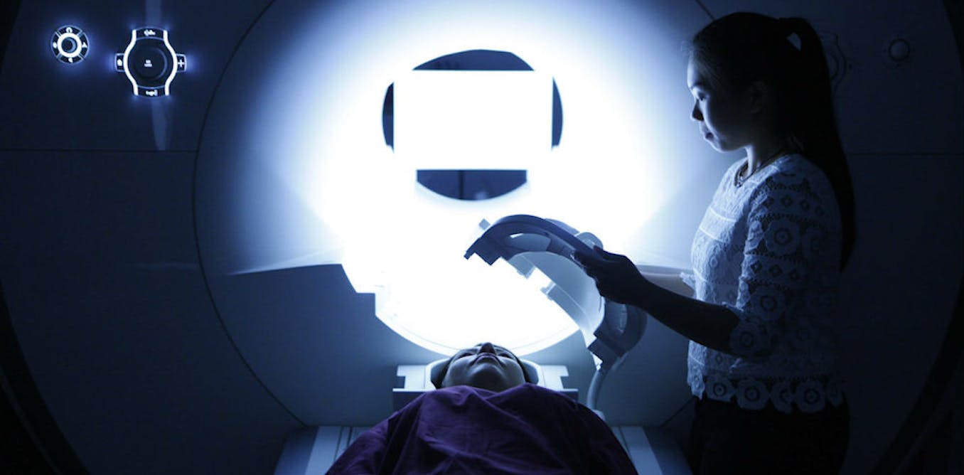
About 8,000 people in the UK are living with Huntington’s disease, a devastating inherited condition that gradually affects movement, thinking and mood. Now, an advanced type of MRI scan could give researchers a way to estimate the cellular damage it causes in living people, which could eventually help show whether treatments are working. New research from my colleagues and I found that the technique can detect abnormalities in the brains of people living with Huntington’s that match those previously identified by examining brain tissue after death. Huntington’s disease is an inherited condition caused by a faulty gene. Its effects usually begin between the ages of 30 and 50. There is currently no cure, although new cell and gene therapies are being developed and tested. One of the main things that happens in Huntington’s is the loss of neuronal cells in the striatum, a part of the basal ganglia deep within the brain. These structures are important for controlling movement and other functions. As cells are lost, the brain tissue in these regions shrinks. We can see this shrinkage using conventional MRI. But a standard brain scan tells us relatively little about what is happening inside the tissue at a cellular level. That’s where our approach comes in. We used a technique called “soma and neurite density imaging”, or Sandi, to analyse diffusion MRI scans. Diffusion MRI detects how water moves through brain tissue. Because that movement is affected by the structures around the water, we can use it to make indirect estimates of properties such as the apparent size and density of cell bodies. This means we can get indirect estimates of the structures within the tissue itself. For our study, we analysed scans from 56 people with Huntington’s disease and 57 healthy volunteers of a similar age and sex. The participants were scanned using a strong-gradient MRI scanner. We focused on the basal ganglia, where we expected to see the effects of Huntington’s disease. We also looked at the thalamus, a nearby brain region that is relatively spared during the early stages of the disease, as a comparison. The results showed a clear difference. In the basal ganglia of people with Huntington’s disease, we found lower estimates of apparent cell-body density, larger estimates of apparent cell-body size and more space between cells than in healthy volunteers. We didn’t see the same pattern in the thalamus. These findings are particularly interesting because they resemble what has previously been seen in brain tissue after death. Postmortem studies have shown that Huntington’s causes a specific type of striatal neurons – the cells that send signals around the brain and nervous system – to be lost. At the same time, glial cells, which normally support and protect neurons, change in response to the damage. They become larger and more active, altering the environment around the remaining neuronal cells. Neurons and glial cells inside the human brain. Kateryna Kon/Shutterstock Our results suggest that Sandi may indirectly capture these biological changes in the living brain. The MRI measurements were also associated with the severity of Huntington’s disease and with poorer performance on finger-tapping tests, which are commonly used to assess motor control. In some regions of the striatum, our estimates of apparent cell-body size and density, together with a person’s age, explained up to 63% of the observed brain shrinkage. This suggests that the measurements may be capturing some of the biological processes driving the loss of brain tissue, and could eventually be important for the development of treatments. Future treatments New cell and gene therapies for Huntington’s disease are being investigated, but researchers need ways of measuring what is happening inside the brain of a living person. If a treatment is designed to protect brain cells, we need to be able to measure whether that is actually happening. Sandi could potentially provide such a measure. It might allow us to indirectly track cellular changes non-invasively and, in future clinical trials, help determine whether a treatment is altering the underlying disease process. But we are not quite there yet. Our study was a snapshot, comparing people with Huntington’s disease and healthy volunteers at a single point in time. This does not tell us whether Sandi can track disease progression, or whether Sandi can reliably detect changes caused by a treatment. We now need larger studies that follow people over a period of time to establish whether these measurements can track the progression of Huntington’s disease. The technique will also need to be adapted and validated for the MRI scanners routinely used in hospitals. Sandi could give us something we currently lack: a way to look beyond brain shrinkage and gain an insight into the cellular changes taking place inside the living brain. And because the loss of brain cells is also a feature of more common conditions such as Parkinson’s and Alzheimer’s disease, the approach may have applications beyond Huntington’s.
New MRI method could help track Huntington’s disease and its treatments
Full Article
Original Source
Read the full article at Theconversation →KhanList aggregates and links to publicly available news content. We do not host full articles from third-party sources. Always verify important information with original sources.