
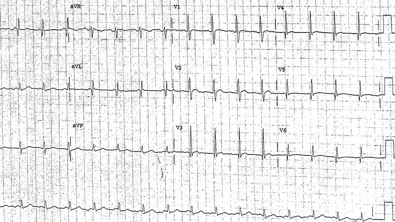
A 20-year-old patient with known proximal muscle weakness and difficulty walking presents to a neurologist for a routine follow-up. A 12-lead ECG is obtained, which prompts a consultation with a cardiologist.Figure 1. The correct diagnosis is sinus tachycardia, short PR interval, and pattern consistent with Duchenne muscular dystrophy (Figure 2).Figure 2.DiscussionThe rhythm is regular at a rate of 100 beats/min. A P wave occurs before each QRS complex (+), with a stable but short PR interval (0.12 sec). The PR interval is shorter than what would be expected for a sinus tachycardia of 100 beats/min and likely represents enhanced atrioventricular (AV) nodal conduction. This may be caused by a rapidly conducting (slick) AV node or a bypass tract of the AV node. As the QRS complex duration is normal (0.08 sec), indicating conduction to the ventricle via the normal His-Purkinje system, this would be considered a Lown-Ganong-Levine (LGL) pattern. LGL pattern is caused by the bundle of James, an accessory pathway that bypasses conduction through the AV node. The QRS complex duration is normal, with a normal axis between 0° and +90° (positive QRS complex in leads I and aVF). However, the QRS complex morphology is abnormal with prominent (although not very wide) Q waves in leads I, aVL, and V4-V6 (^), suggesting an old lateral-wall myocardial infarction. There is also a tall R wave in lead V1 (←). Causes of a tall R wave in lead V1 include: Right ventricular hypertrophy (usually associated with a right axis and P-pulmonale)Posterior wall MI. This is often associated with an inferior wall MI, due to a lesion in the left circumflex or obtuse marginalDextrocardia (with reverse R wave progression and what looks like R-L arm lead switch with a right axis and also negative P and T waves in leads I and aVL)Right sided leads (with reverse R wave progression)V1-V3 lead switchWolff-Parkinson-White pattern (with short PR interval and delta wave)Significant counterclockwise rotation of the electrical axis in the horizontal planeHypertrophic cardiomyopathyDuchenne muscular dystrophy (with a posterolateral infarction pattern)Because this ECG shows a posterolateral infarction pattern, it indicates Duchenne muscular dystrophy. This pattern may also be seen in Becker muscular dystrophy. Both conditions are often associated with a cardiomyopathy due to myocardial fibrosis, which begins in the posterolateral part of the left ventricle.Philip Podrid, MD, is an electrophysiologist, a professor of medicine and pharmacology at Boston University School of Medicine, and a lecturer in medicine at Harvard Medical School. Although retired from clinical practice, he continues to teach clinical cardiology and especially ECGs to medical students, house staff, and cardiology fellows at many major teaching hospitals in Massachusetts. In his limited free time he enjoys photography, music, and reading.
ECG Challenge: Neuro Patient With Difficulty Walking
Full Article
Original Source
Read the full article at Medscape →KhanList aggregates and links to publicly available news content. We do not host full articles from third-party sources. Always verify important information with original sources.