
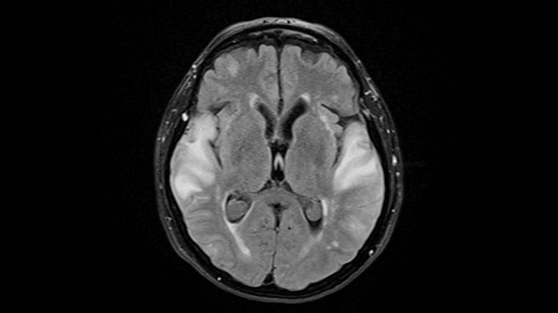
A known genetic risk factor for amyloid-related imaging abnormalities (ARIA) associated with the antiamyloid lecanemab may offer little help in predicting who will actually develop the potentially serious complication, new real-world data suggest.APOE epsilon 4 was associated with a greater likelihood of ARIA — particularly among homozygous carriers — but it did not reliably identify who would go on to develop the complication.In a single-center retrospective study of 230 patients treated with lecanemab, 24.3% developed ARIA, with most moderate-to-severe cases emerging within the first 24 weeks of treatment.Risk was higher among those with two copies of APOE epsilon 4: Homozygotes had nearly fivefold higher odds of ARIA with edema (ARIA-E) and nearly fourfold higher odds of ARIA with microhemorrhages (ARIA-MH) than with noncarriers. However, APOE epsilon 4 and other clinical, imaging, and cerebrospinal fluid (CSF) markers could not reliably predict which patients would develop ARIA.“APOE cannot predict who will develop ARIA at the individual level,” lead investigator Andy J. Liu, MD, MS, associate professor of neurology and pathology at Duke University School of Medicine in Durham, North Carolina, told Medscape Medical News. “It is still useful information because we commonly counsel patients on their potential risk.”The study was published online on August 7 in Neurology Open Access.Real-World SafetyIn 2023, the FDA approved lecanemab for the treatment of amyloid-positive mild cognitive impairment (MCI) or mild Alzheimer’s disease dementia.In the pivotal CLARITY AD trial, lecanemab slowed clinical decline but was also associated with an increased risk for ARIA.Although clinical trials established ARIA as an important safety concern with lecanemab, questions remain about how frequently and when these events occur in routine clinical practice and whether those at greatest risk can be identified before treatment. The retrospective study was designed primarily to assess the incidence and timing of ARIA in real-world lecanemab use, including differences by diagnosis, APOE epsilon 4 genotype, sex, and race.Secondary aims included identifying baseline predictors of ARIA, examining whether ARIA was associated with changes in cognition, and assessing serious adverse events and mortality.The study included 230 patients (mean age, 73.6 years; women, 50.9%; White, 92.4%) treated at a single academic center between May 2023 and June 2025. Within the cohort, 68% of participants had MCI, and 67.4% had carried at least one APOE epsilon 4 allele (55.7% heterozygous and 11.7% homozygous). Amyloid pathology was confirmed via CSF biomarkers in 68% of patients and florbetapir PET in 32% of patients.Participants underwent standardized MRI surveillance, including susceptibility-weighted imaging to detect microhemorrhages and superficial siderosis. MRI was performed after the fourth, sixth, and 13th infusions, with additional monthly imaging for those who developed ARIA. The center later added MRI after the second infusion in accordance with updated FDA recommendations.They also underwent APOE genotyping and, when available, CSF biomarker testing. Those who took anticoagulants or had at least five cerebral microhemorrhages or superficial siderosis (ARIA-SS) at baseline MRI were excluded.Given concerns about hemorrhagic complications with lecanemab, four participants with atrial fibrillation underwent left atrial appendage occlusion before treatment, allowing them to discontinue anticoagulation while maintaining stroke prevention.Although APOE epsilon 4 homozygotes were not excluded because of their higher ARIA risk, the center provided individualized counseling on the potential risks and benefits of treatment.ARIA Common, Difficult to PredictOverall, 56 patients developed ARIA, with some experiencing more than one subtype. ARIA-MH was the most common, affecting 49 patients, followed by ARIA-E in 22 and ARIA-SS in nine. ARIA-E was observed throughout treatment, with incidence peaking at approximately weeks 10 and 25. Most ARIA-E and ARIA-SS events emerged within the first 17 weeks, whereas ARIA-MH events occurred over a longer period, extending to approximately 27 weeks.Moderate-to-severe ARIA was most common within the first 24 weeks of treatment, although milder events emerged later. Among those with follow-up imaging documenting resolution, ARIA resolved within approximately 3-20 weeks.APOE epsilon 4 homozygosity was strongly associated with ARIA risk. Compared with noncarriers, those with two epsilon 4 alleles had higher odds of ARIA-E (odds ratio [OR], 4.81; P = .014) and ARIA-MH (OR, 3.76; P = .008). In contrast, carrying a single epsilon 4 allele was not associated with a statistically significant increase in either subtype (OR, 1.19 for ARIA-E; OR, 1.10 for ARIA-MH). Despite these associations, APOE epsilon 4 status could not reliably predict who would develop ARIA.The multivariable model, which incorporated age, diagnosis, sex, race, APOE epsilon 4 allele count, CSF p-tau181/amyloid beta 42 ratio, and Fazekas score, had limited predictive accuracy, with an area under the curve of 0.58. Individual markers showed high specificity but low sensitivity, limiting their ability to identify patients who will develop ARIA.Stable Cognition After ARIAOver 12 months, Montreal Cognitive Assessment scores declined by 0.107 points per month (P = .002), but the rate of decline did not differ significantly between those with and without ARIA (P = .364).Mini-Mental State Examination (MMSE) scores showed a nonsignificant decline of 0.052 points and also no significant difference between the groups. However, patients with ARIA experienced a transient drop in MMSE at 6 months that appeared to reverse by 12 months.“It is reassuring for clinicians to know and counsel patients that despite experiencing ARIA, cognitive abilities 12 months later show no significant changes,” Liu said.Despite encouraging cognitive findings, lecanemab’s safety profile highlighted challenges of using the drug in routine clinical practice.Overall, there were a total of 72 clinically significant adverse events, including 22 falls, 10 strokes, seven severe infusion reactions, and three seizures. About 27 patients required hospitalization, and five deaths occurred, including two considered related to lecanemab. Treatment was discontinued in 49 patients, including 19 because of ARIA.The study had several limitations, including its retrospective, single-center design, small sample size, lack of a control group, and predominantly White cohort. The exclusion of patients taking anticoagulants further limits the generalizability of the safety findings. Longer follow-up is also needed to determine whether cognitive stability after ARIA is sustained.From Risk Factors to Personalized PredictionThe distinction between a risk factor and an individual predictor is important when interpreting the study’s findings, said Lawrence S. Honig, MD, PhD, professor of neurology at Columbia University Irving Medical Center in New York City, who was not involved in the study.“Even in those with the most unfavorable genetic risk, namely APOE epsilon 4 homozygotes, 76% in this study did not get ARIA-E of any severity,” Honig told Medscape Medical News.Given the limitations of current risk models, Honig said the goal should be to improve individualized risk assessment while recognizing that treatment decisions will always involve some uncertainty.“It is very reasonable to strive for patients and practitioners to have a better, personally individualized assessment of risk of ARIA,” he said. “Individuals should be able to decide themselves how the risk of disease-slowing treatments, which is always statistical and uncertain, impacts their decisions on considering such therapy.”An accompanying editorial by Marco E. Egle, MD, PhD, and Rebecca F. Gottesman, MD, PhD, underscored the challenge of translating population-level risk into individualized predictions, arguing that future approaches will need to account for multiple interacting factors.“The inability of this genetic marker, or any other commonly available baseline characteristic, to reliably predict an ARIA event” is a key limitation of current risk models.Rather than considering individual factors in isolation, Egle and Gottesman suggest that future risk models could use machine learning to identify combinations of cerebrovascular, cardiovascular, and metabolic factors that increase ARIA susceptibility, integrating these data with neuroimaging and fluid biomarkers.“Integrating these multimorbidity profiles with neuroimaging and fluid biomarkers presents a promising path forward,” they concluded.Disclosure information for study authors is available in the original study publication. Honig reported receiving research funding or consulting fees from the National Institutes of Health (NIH)/National Institute of Neurological Disorders and Stroke, NIH/National Institute on Aging, Eisai, Genentech/Roche, Biogen, Bristol Myers Squibb, Eli Lilly, and UCB, among others.
APOE epsilon 4 Flags ARIA Risk but Falls Short as Predictor
Full Article
Original Source
Read the full article at Medscape →KhanList aggregates and links to publicly available news content. We do not host full articles from third-party sources. Always verify important information with original sources.