
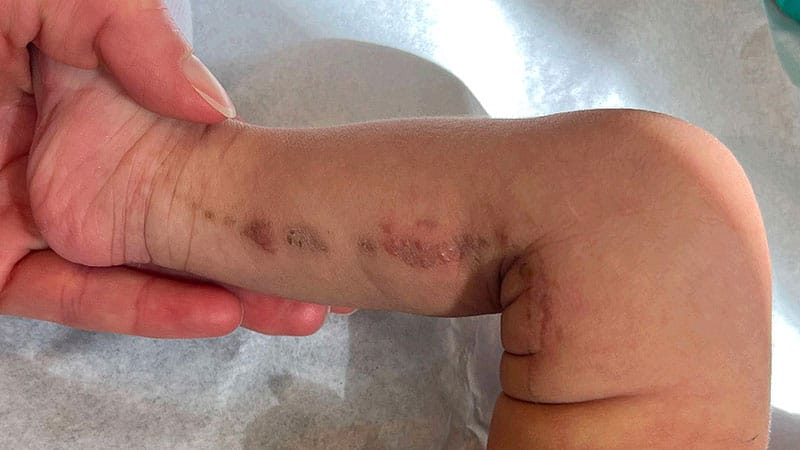
A 5-week-old male presents to the dermatology clinic for evaluation of a blistering rash on his right leg. The blisters have been present since birth and have darkened over time. His parents do not believe the area is associated with any pain or pruritus and deny any discharge or bleeding. Examination revealed pink inflammatory papules and vesicles with areas of brown scale in a linear distribution extending from just above the right medial malleolus to the right inguinal crease, with no involvement of the right foot. The child is otherwise well. His mother reports that prenatal labs, genetic testing during pregnancy, and newborn screenings were all within normal limits. The child has no history of recent illness, no sick contacts, and no family members with similar lesions.The correct diagnosis is incontinentia pigmenti.DiscussionThis patient has a linear distribution of vesicles extending from the right medial malleolus to the right inguinal fold. Given the linearity of the vesicles, their presentation at birth with hyperpigmentation over time, and the lack of associated pain or signs of infection, the lesion is most consistent with incontinentia pigmenti (IP). This case is unusual in that the affected child is male.IP is a rare genetic condition of X-linked inheritance characterized by abnormalities of the skin, teeth, eyes, and central nervous system (CNS). It is named after its characteristic brown-blue-gray discoloration that is histologically correlated with dermal pigmentation (as compared to epidermal pigmentation), referred to as incontinent pigment. IP is caused by mutations in the IKBKG gene, which is involved in the regulation of cell division and apoptosis and important in NF-NF-κB signaling, making cells with the mutation more prone to death. This cell death can cause skin, hair, nail, and teeth disfiguration or loss; eye defects or blindness; and neurologic complications such as seizures, intellectual disability, spastic paralysis, and cerebral atrophy. However, progressive skin rashes are the main feature of IP; more severe disease with eye or CNS involvement only occurs in approximately 30% of patients.Savannah Bush, BSBecause IP is X-linked dominant, only one copy of the abnormal gene on the X chromosome will cause disease. Females with variants of the IKBKG gene on one X chromosome have a normal gene on their other X chromosome, whereas males do not, given their XY chromosomal makeup. As a result, most males who inherit the mutated gene die in utero. Just over 100 male cases of IP have been published. Somatic mosaicism is the most likely explanation for these cases, with postzygotic mutations (mutations that occur after fertilization and are only passed to daughter cells of the mutated somatic cell) leaving some cells without the variant unaffected. Other cases may be associated with Klinefelter syndrome (47,XXY), with the second X chromosome “balancing” the abnormal gene. The defining skin lesions of IP are present at birth or develop within the first weeks of life and classically progress through four stages. Stage 1 lesions are vesicular, presenting with red, blister-like lesions grouped in a line, most commonly on the extremities, scalp, or trunk. Stage 2 lesions are verrucous, with lesions often darkening in color and the formation of thick scabs or crusts on top of previous blisters. Stage 3 occurs once scabbing resolves and hyperpigmented patches remain. Stage 4 may occur once the patient enters adolescence or adulthood, with atrophy or hypopigmentation of prior lesions.Lawrence F. Eichenfield, MDThere is no treatment for IP. Management includes prevention of infection associated with skin lesions and monitoring for additional disease manifestations, ophthalmologic screening, and dental care.The differential diagnosis for IP includes lichen striatus, epidermal nevus, Herpes simplex virus ( HSV) infection, and impetigo. Lichen striatus is unlikely in this patient as it is acquired and noncongenital. The lesions of lichen striatus are small, 1-4 mm lichenoid papules in a linear configuration, but not vesicular. Lichen striatus is self-limited with resolution in months to years. Epidermal nevi can also present at birth and may also be associated with somatic mutations. However, epidermal nevi are hamartomatous and characterized by focal skin-colored, tan, or brownish plaques, often in a linear pattern. They do not present with vesicles.HSV infection may be considered in the differential diagnosis of IP, as it presents with vesicles, although in HSV infection, the lesions are not grouped linearly. Impetigo can also cause vesicular lesions; however, these lesions are rarely linear, tend to weep, and are associated with honey-colored crusting.Recommended ReadingPacheco T, Levy M, Collyer JC, et al. Incontinentia pigmenti in male patients. J Am Acad Dermatol. 2006;55:251-255. SourceJabbari A, Ralston J, Schaffer JV. Incontinentia pigmenti. Dermatol Online J. 2010;16:9. Source
A 5-Week-Old Male Presents With Blisters on His Right Leg
Full Article
Original Source
Read the full article at Medscape →KhanList aggregates and links to publicly available news content. We do not host full articles from third-party sources. Always verify important information with original sources.